Researchers identify dormant progenitor cells in skeletal muscle that migrate to fracture sites and become bone-forming cells
CHINA, August 14, 2026 /EINPresswire.com/ — Bone healing, an intricate process involving multiple cell populations, relies not only on existing bone-forming cells but also on dormant progenitor cells outside the skeleton. A study published in Bone Research reveals that fibroadipogenic progenitors (FAPs) in skeletal muscle become bone-forming cells after injury, contributing to fracture repair and abnormal bone growth. The findings identify these cells as promising therapeutic targets for enhancing fracture healing while preventing unwanted bone formation.
Bone healing is a complex process that depends on the coordinated activity of many different cell types. While bone-forming cells called osteoblasts are known to rebuild damaged tissue, researchers have increasingly found that some cells outside the skeleton remain dormant under normal conditions but can acquire bone-forming functions after injury. Given the diversity of cell populations in the musculoskeletal system, the precise role of individual cell types remain unexplored.
Against this backdrop, a study published online in Volume 14 of the journal Bone Research on July 06, 2026, revealed an unexpected role of muscle-resident fibroadipogenic progenitors (FAPs), together with a smaller population of superficial periosteal cells, in repairing fractured bones. The study was led by Dr. Ugur M. Ayturk and colleagues from the Skeletal Health and Orthopedic Research Program, USA.
FAPs reside in skeletal muscle, while superficial periosteal cells are found in the thin connective tissue called periosteum, covering the outer surface of bones. Although these cells normally remain inactive, they are recruited following injury to help repair fractured bones.
Dr. Ayturk says, “Our findings show that extra-skeletal cells, particularly FAPs, are recruited to help repair bone fractures and could represent a promising therapeutic target to enhance fracture healing.”
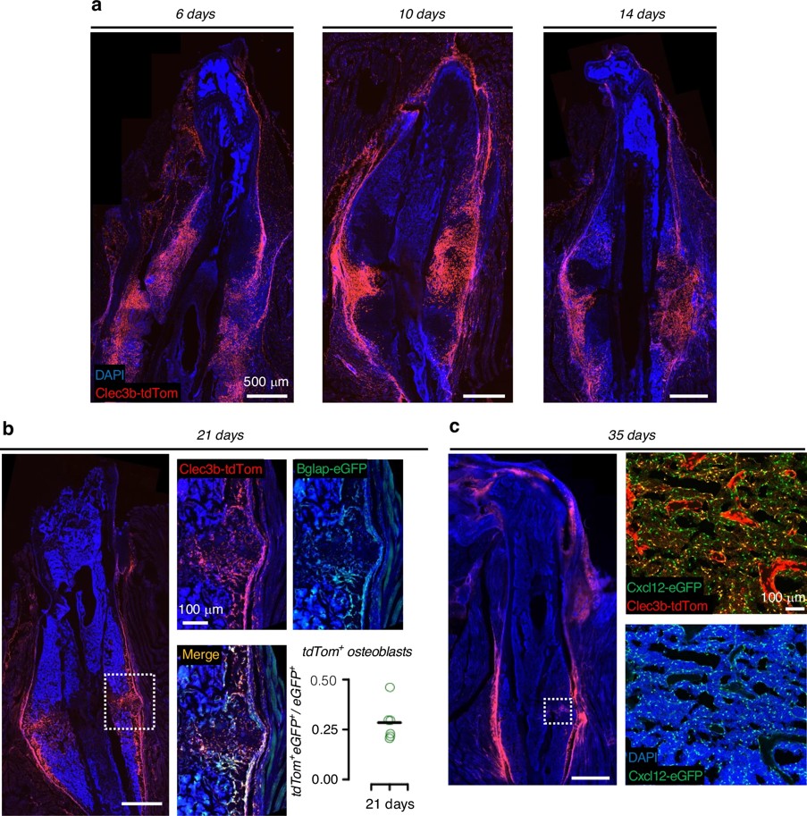
To study these cells, the researchers found Clec3b expression as a highly specific marker for these normally dormant progenitor cells. They engineered a mouse model in which they labelled cells expressing Clec3b with a fluorescent tag, allowing them to track their location under normal conditions and their response after injury.
During normal bone growth, they found that these cells remained in muscle and the superficial periosteum and they never migrated into bone or differentiated into osteoblasts. Following bone fractures, however, these cells rapidly migrated to the injury site, where many differentiated into osteoblasts that produced new bone and aiding in healing process. Within three weeks, the researchers found that about 28% of the osteoblasts in the healing callus originated from Clec3b-lineage cells. Some of these cells also became bone marrow stromal cells, which helped rebuild the supportive environment inside the bone. Notably, the cells stopped expressing Clec3b as they differentiated, indicating that the marker is associated with their dormant progenitor state.
Single-cell RNA sequencing confirmed this transition, showing that dormant Clec3b-lineage cells gave rise to new populations with the molecular characteristics of bone marrow stromal cells and osteoblasts after fracture.
Further, the researchers investigated the origin of these bone-forming cells. Although similar cells are also found in the periosteum, their experiments showed that skeletal muscle is the main source. Even after the periosteum was surgically removed before injury, Clec3b-positive cells still reached the fracture site and developed into bone-forming cells. Bone grafts that included surrounding muscle generated substantially more Clec3b-lineage bone-forming cells than grafts without muscle, indicating that skeletal muscle is the primary source of these regenerative cells.
The cells were also found to contribute to heterotopic ossification, a condition in which bone forms in muscles and other soft tissues after injury. In mouse models, Clec3b-lineage cells differentiated into cartilage- and bone-forming cells, becoming a major source of this abnormal bone. “When we blocked a key pathway required for bone formation or depleted these cells, both fracture healing and abnormal bone growth were significantly reduced. This finding highlights their important role in both processes,”shares Dr. Ayturk.
The researchers believe these cells could become promising therapeutic targets. Activating them may enhance fracture healing, while limiting their bone-forming activity could help prevent unwanted bone growth following serious injuries.
***
Reference
Title of original paper: Clec3b+ extraskeletal cells regulate fracture healing and heterotopic ossification
Journal: Bone Research
DOI: 10.1038/s41413-026-00532-6
About Bone Research
Bone Research was founded in 2013. As a new English-language periodical, Bone Research focuses on basic and clinical aspects of bone biology, pathophysiology and regeneration, and supports the foremost discoveries resulting from basic investigations and clinical research related to bone. The aim of the journal is to foster the worldwide dissemination of research in bone-related physiology, pathology, diseases and treatment.
Website: https://www.nature.com/boneres/
About Dr. Ugur Ayturk from Skeletal Health and Orthopedic Research Program, USA
Dr. Ugur Ayturk is an Assistant Scientist in the Musculoskeletal Integrity Program at Hospital for Special Surgery (HSS) and an Assistant Professor of Physiology and Biophysics in Orthopaedic Surgery at Weill Cornell Medicine. He joined HSS in 2017 after serving as an Instructor in Orthopaedic Surgery at Boston Children’s Hospital and Harvard Medical School. Dr. Ayturk earned his Ph.D. from Colorado State University and completed postdoctoral training at Boston Children’s Hospital. His research focuses on understanding how osteoblasts develop from mesenchymal progenitor cells and how these cells regulate bone formation, repair, and regeneration.
Funding information
This study was supported by grants from the S&L Marx Foundation, Giuliani Foundation, and Stavros Niarchos Complex Joint Reconstruction Center at Hospital for Special Surgery, with additional fellowship support provided by the Francis Glorieux Research Fellowship from the Canadian Osteogenesis Imperfecta Society.
Yini Bao
Bone Research Editorial Office
+86 2885546461
br@scu.edu.cn
Visit us on social media:
X
Legal Disclaimer:
EIN Presswire provides this news content “as is” without warranty of any kind. We do not accept any responsibility or liability
for the accuracy, content, images, videos, licenses, completeness, legality, or reliability of the information contained in this
article. If you have any complaints or copyright issues related to this article, kindly contact the author above.

Media gallery